
B-Bridgeの事業
B-Bridgeが日本とシリコンバレーの架け橋に
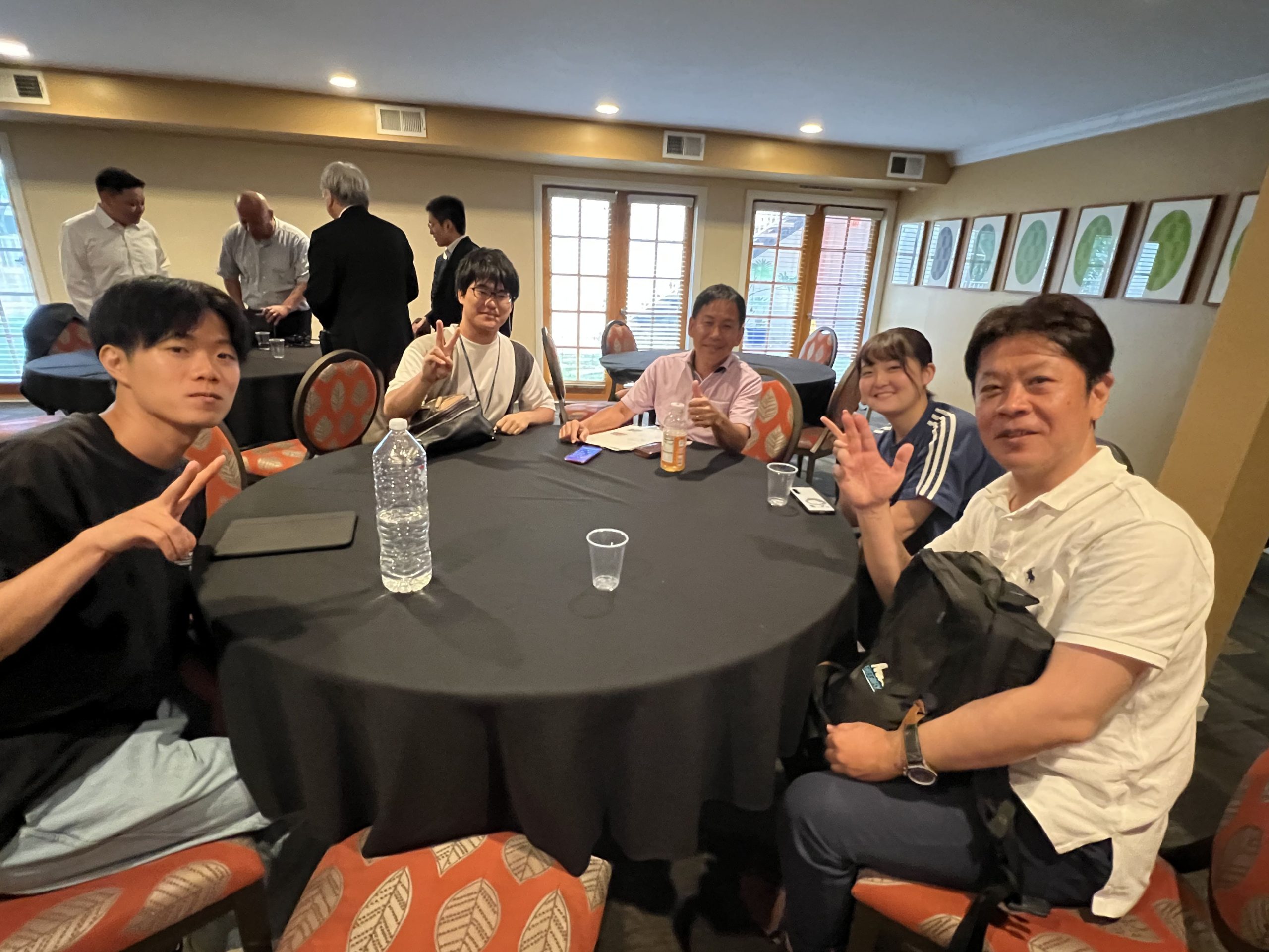
B-Bridgeは日本とシリコンバレーの架け橋となり、様々な事業の橋渡しをしています。日本の事業を海外展開したい、グローバル人材を育成したい、アスリートやアーティストのチャレンジの場を探したい。そんなチャレンジができる場がシリコンバレーです。そのための支援をB-Bridgeは行なっています。
-

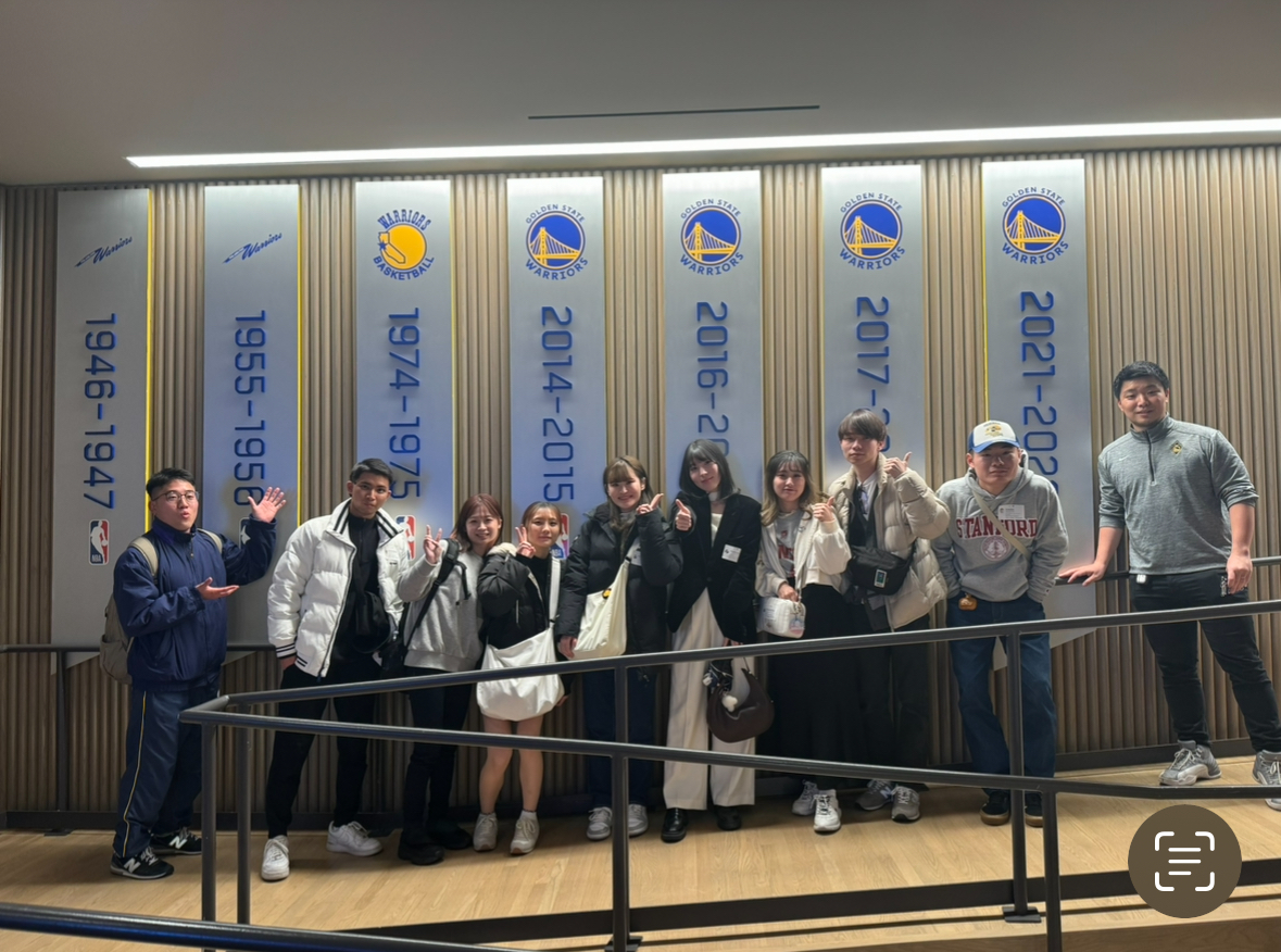
ビジネス支援 Business Incubation
シリコンバレーでのチャレンジをB-Bridgeが全力でサポートします。
-
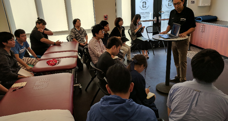
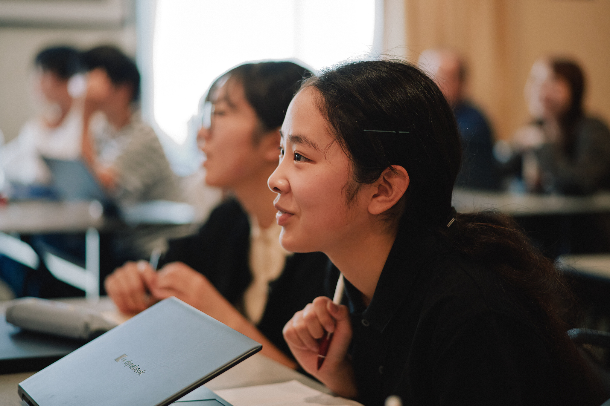
研修・人材育成 Education
グローバルに通用する人材育成や最先端のシリコンバレーを知る研修を行います。
-

オンラインサロン Online Salon
様々な強みを持ったプロたちのコンテンツを届けるコミュニティを形成しています。
-
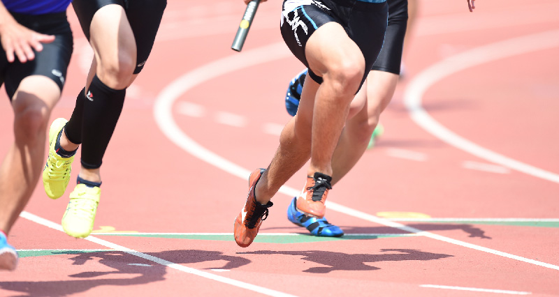
アスリートマネジメント Athlete Management
アスリートのデュアルキャリア形成をバックアップします。
-

アーティストマネジメント Artist Management
音楽家・芸術家が長くアート活動できるよう、支援します。
-

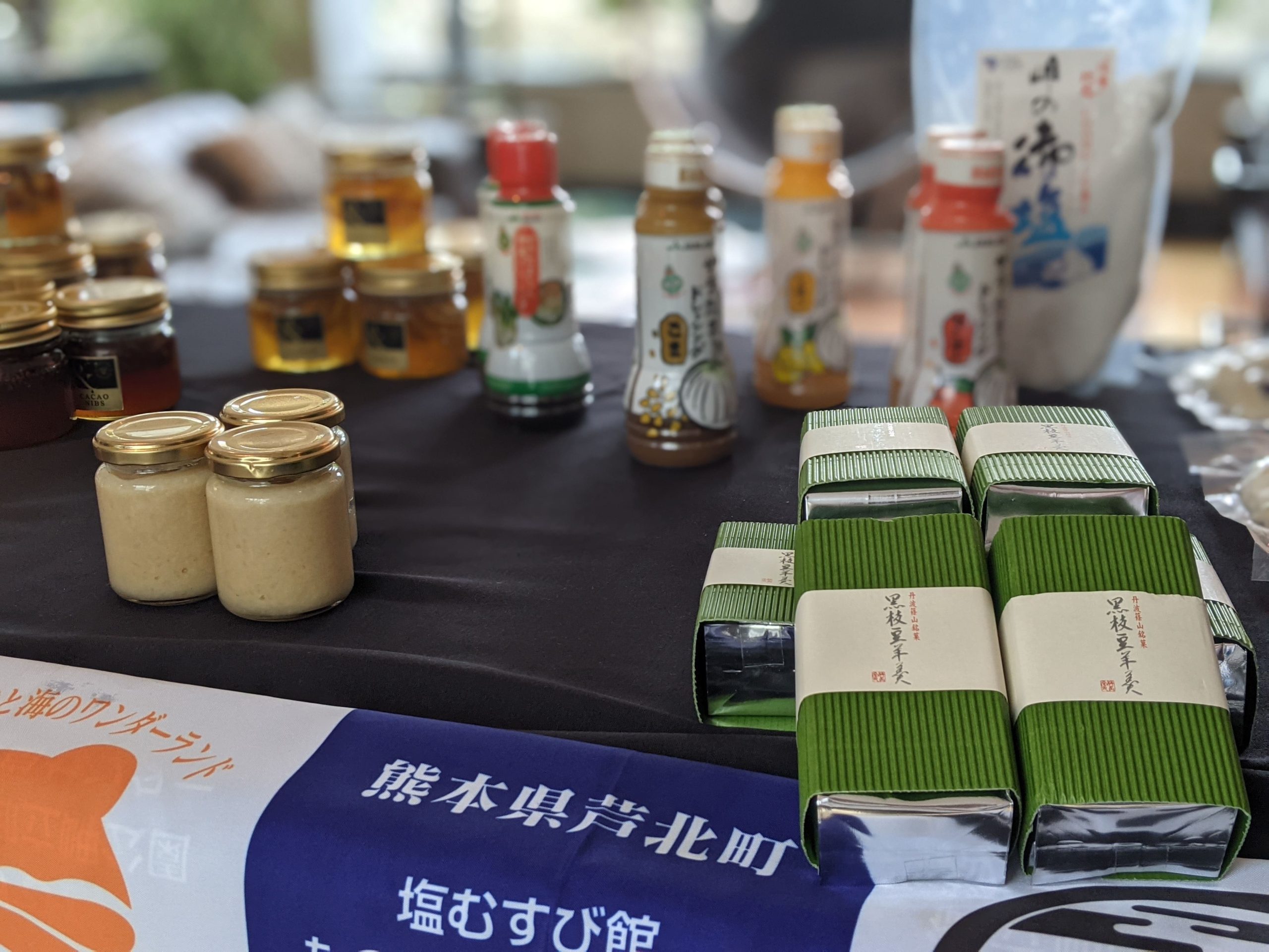
地方創生支援 Region Revitalization
地方の事業をスケールさせるためのヒントをシリコンバレーで見つける為の支援です。
コラム・ブログ
B-Bridgeがお届けする最新情報。
お問い合わせ
B-Bridgeへのご相談、事業についてお問い合わせのある方はこちらからお気軽にご相談ください
フォームからのお問い合わせ
問い合わせフォームからお気軽にご相談いただけます。
受付は24時間しております。